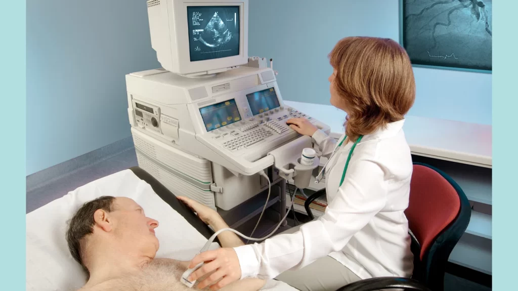
Echocardiography is an important instrument used in contemporary medicine to understand the condition of the heart. We will discuss what echocardiogram Upper East Side is, how it functions, why it is important, and what to anticipate if you ever need one in this blog post.
What is an echocardiogram?
A medical test called an echocardiogram, sometimes referred to as an “echo,” uses sound waves to produce live images of the heart. It is non-invasive, painless, and extremely informative. Essentially, it is an ultrasound of the heart.
Why is echocardiography important?
- Heart Structure: Echocardiograms make the chambers, valves, walls, and blood arteries of the heart visible, enabling medical professionals to evaluate both the structure and function of the organ.
- Functionality: It offers important information regarding the efficiency of blood pumping in the heart, which is critical for determining heart function as a whole.
- Diagnosis: Echocardiography is used to identify and track the number of cardiac problems such as cardiomyopathies, congenital heart abnormalities, murmurs, and heart valve illnesses.
- Treatment Guidance: They help doctors choose the best course of action, including whether to perform surgery or prescribe medicine.
How Does an Echocardiogram function?
An echocardiogram produces precise images of the heart using high-frequency sound waves (ultrasound). What normally occurs throughout the operation is as follows:
- Gel Application: To enhance sound wave conduction, a technician administers a specific gel on the chest.
- Transducer Positioning: The technician will next insert a little gadget on the chest, which is known as a transducer. Sound waves are emitted by the transducer, which captures the echoes as they return.
- Image Generation: The transducer sends these echoes to a computer, which displays real-time images of the heart on a screen as it moves across the chest.
- Evaluation: To determine the anatomy and function of the heart, a cardiologist or technician will evaluate the images.
Types of Echocardiograms
There are different types of echocardiograms, including
- Transthoracic echocardiogram (TTE): It is the most frequent type and is performed via the chest wall.
- Transesophageal echocardiogram (TEE): A probe was inserted into the esophagus through the mouth to obtain a closer look at the heart.
- Stress echocardiogram: This test measures how the heart reacts to stress and is performed both before and after exercise or medication.
Echocardiography is an important diagnostic tool in cardiology. They allow medical professionals to make knowledgeable choices regarding your heart health because they offer in-depth insights into the anatomy and operation of your heart. If your doctor advises getting an echocardiography, know that it is a painless, safe technique that can provide vital details regarding your cardiovascular health. Your heart appreciates this.
About Author
You may also like
-
CBT Coaching for Anxiety: Empowering Change Through Thought and Action
-
Why Diabetic Retinopathy Screening Shouldn’t Be Ignored
-
Top Medical Transcription Software for Accurate Healthcare Documentation
-
Targeted Solutions for Every Skin Concern: From Acne Scar Treatment to Botox Treatment Singapore
-
The Science Behind Drinkable Collagen: What Studies Really Say


